Module III:
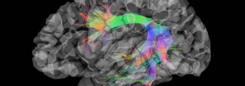
Development of Neuroimaging and Modeling Methods
Cutting-edge imaging, neurostimulation, data analysis, and modeling play a crucial role in the success of cognitive and human neuroscience. They offer non-invasive access to brain structure and function and even selective modulation of brain activity. Modeling provides quantitative predictions for experiments and facilitates the interpretation of experimental results within a conceptual framework. This structures research approaches and methods.
Thus, training in the different methods of neuroimaging, neurostimulation, data analysis, and modelling, and their development is one of the pillars of the IMPRS CoNI. The advanced training courses go far beyond traditional programs as the collection of methods taught are only available at a few sites around the world. The IMPRS CoNI covers all major imaging and neurostimulation methods and their appropriate use.
These include:
- magnetic resonance imaging (MRI)
- electroencephalography (EEG)
- magnetoencephalography (MEG)
- positron emission tomography (PET)-MRI
- near-infrared spectros-copy (NIRS)
- transcranial magnetic stimulation (TMS),
- direct current stimulation (tDCS).
Research focus are Spiking Neural Networks, specially neurological plausible learning algorithms as well as the incorporation of dynamcs within synaptic efficiency towards SNNs.
more
Computational modelling of brain networks, neural mass modelling. | Biophysical modelling of EEG/MEG, in particular source reconstruction.
more
Analysis and modeling of MEG/EEG data to localize and identify brainnetworks
more
Methods for MRI and MRS including pulse sequences, RF hardware and image processing.
more
The key goal of the Computational Machinery of Cognition (CMC) lab is to understand the computations that support goal-driven behavior, and also the multi-scale machinery (neurons, …, large-scale networks) that implements these computations.
more
How can a network of cells perceive, represent, and make sense of the world around it? Experimental techniques such as high-resolution, multi-modal magnetic resonance imaging, light-sheet microscopy, or single-cell sequencing provide unprecedented insights into the structures and processes of neural systems and life in general. However, these methods yield high-dimensional data with intricate structures and dependencies that can be challenging to interpret.
more
Multimodal visualization of brain imaging data, like MRI, DT-MRI, HARDI, fMRI, EEG, MEG
more
Our vision is to develop and apply functional microstructure imaging and in-vivo histology using magnetic resonance imaging (MRI) as novel non-invasive MRI methods to reliably characterize the detailed functional and anatomical microstructure of the human brain.
more
